The goal of this course is to become familiar with the good practices for the analysis of small geological objects of microscopic size. These objects, such as fluid, vitreous or mineral inclusions in minerals for example, are frequent in Earth Sciences and often carry rich and key information. On such small objects, analyzes shall be carried out in situ, before perhaps sacrificing them for a final analysis requiring extraction. The example of materials under extreme pressure and temperature conditions, mandatory to model the physico-chemical properties of the interior of the Earth and planets will also be discussed.
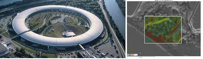
Left : European Synchrotron Radiation Facility. Right : EDS mapping of a synthetic sample produced at the temperature and pressure of a subduction zone
The main techniques of in situ microanalysis, to gain chemical and physical information, will be presented – approximately 2 hours lecture, and then practically implemented when available in our partner laboratories – lab work of 3 to 4 hours. Otherwise, data available in the instructors' portfolio will be used. in situ tant chimiques que structurales seront présentées – 2 h de cours environ, et mises en œuvre lorsque celles-ci sont présentes dans les laboratoires partenaires – TP de 3 à 4h. Dans le cas contraire, des données disponibles dans le portfolio des enseignants seront utilisées.
Following each lab work, students write an experience report illustrating the relevance and limitations of the technique (personal work estimated at 4 hours/topic).
The class includes a visit to the ESRF (European Synchrotron Research Facility) in Grenoble to raise the awareness of students on the great potential of selected beamlines and more generally large international facilities.
Methods and analyses (the list may be adapted according students’ interests or needs and/or availability of equipment):
Advanced SEM scanning electron microscopy – including EDS, cathodoluminescence, and EBSD measurements;
Transmission electron microscopy TEM and possibly Focussed Ion Beam (FIB) or tomographic atomic probe;